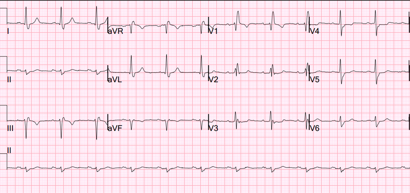

Development of microemboli due to stagnant blood flow from the atria.This causes chaotic myocardial responses that may diminish both the pre-load and effectiveness of the cardiac contraction. Formerly known as chronic or permanent atrial fibrillationĪtrial fibrillation is when multiple electrical impulses are being generated in the atria at the same time.Persistent atrial fibrillation that lasts longer than one year.Long-Standing Persistent Atrial Fibrillation.Episodes that last less than a week but are only stopped by either pharmacological or electrical cardioversion.May last anywhere from seconds or minutes, to hours or up to a week.Transient episodes that stop on their own.only warfarin should be used with vascular lesions (eg.There are three primary types of atrial fibrillation:.international normalized ratio (INR) target of 2-3.novel anticoagulants contraindicated in renal failure.score of 2 or more use oral anticoagulation.e.g., previous myocardial infarction and peripheral artery disease.Stroke/transient ischemic attack/thromboembolism = 2 point.stroke risk stratification CHA 2DS 2-VASc score.in order to decrease the risk of thromboembolism.in heart failure with reduced ejection fraction.beta blocker preferred in coronary artery disease.treatment is chosen based on patient's comorbidities.β-blocker or nondihydropyridine calcium channel blocker.use calcium channel blocker or titratable beta blocker (esmolol).intravenous β- blockers or nondihydropyridine calcium channel blocker.patients with new onset atrial fibrillation become symptomatic due to rapid ventricular response (except in cases of stroke).associated with alcohol, caffeine, and nicotine use.e.g., chronic obstructive pulmonary disease (COPD).commonly seen in patients with pulmonary disease.hyperthyroidism is a possible cause of atrial fibrillation.
#Paroxysmal atrial fibrillation ecg free
thyroid stimulating hormone (TSH) and free T4 level.transesophageal echocardiogram (TEE) is more sensitive in detecting thrombi in the left atrium.can also assess for valvular and pericardial disease, and peak right ventricular pressure.can assess atrial size and ventricular function, thickness, and size.these patients are hemodynamically stable.Holter monitoring in the outpatient setting.if arrhythmia is not captured on ECG then.bibasilar rales on pulmonary auscultation.in cases of atrial fibrillation leading to heart failure.focal neurological deficit if this results in an embolic stroke.shortness of breath (suggesting heart failure).especially after coronary artery bypass grafting (CABG).alcohol abuse ("holiday heart syndrome").e.g., dilated and hypertrophic cardiomyopathy.


 0 kommentar(er)
